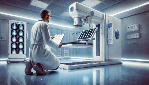
Full Field Digital Mammography Quality Control Checklist
📸
Full Field Digital Mammography Quality Control Checklist
Explore our comprehensive "Full Field Digital Mammography Quality Control Checklist", encompassing hardware checks, image testing, system updates, and detailed reporting.
1
Check digital mammography unit for any physical abnormalities
2
Turn on the mammography unit and warming up
3
Inspect control panel and monitors for any discrepancies
4
Check the process date and time on the monitor for accuracy
5
Perform clinical image review for a selected test image
6
Approval: Clinical Image Review
7
Perform image quality test on the mammography unit
8
Check image receptor resolution and noise
9
Evaluate detector element failures
10
Check grid artifact detection and artifact suppression functions
11
Conduct detector calibration checks
12
Measure contrast to noise ratio for digital mammography images
13
Evaluate image lag and ghosting
14
Check automatic exposure control function
15
Evaluate image display function on the monitor
16
Check system performance trending and problem logs
17
Perform routine mechanical and safety checks
18
Check for system software updates and install them if necessary
19
Approval: System Software Update
20
Document all findings and final assessment in the quality control report
Check digital mammography unit for any physical abnormalities
Inspect the digital mammography unit for any physical abnormalities that may affect its performance. Look for any visible damages, loose connections, or signs of wear and tear. Ensure that all cables and wires are properly connected and secured.
The physical condition of the mammography unit plays a crucial role in ensuring the quality and accuracy of the imaging process. Any abnormalities or damages can affect the results and may require immediate repair or replacement. Regular inspection and maintenance of the unit are necessary to prevent any potential issues and ensure optimal performance.
How to perform: Visually examine the mammography unit, paying attention to the exterior, cables, connectors, and other components. Gently move the unit to check for any loose parts or abnormal sounds. Refer to the manufacturer's guidelines or user manual for specific instructions.
Potential challenges: Some damage or abnormalities may be difficult to detect visually. In such cases, additional diagnostic tests may be required. If any issues are identified, contact the appropriate personnel or IT support to resolve the problem.
Required resources or tools: Visual inspection, knowledge of normal unit appearance and functioning.
Turn on the mammography unit and warming up
Power on the digital mammography unit and allow it to warm up before performing any tests or procedures. This step ensures that the unit is fully functional and ready for use.
Warming up the mammography unit is essential to stabilize its internal components and optimize image quality. It also allows the unit to reach its operating temperature, ensuring accurate and consistent results.
How to perform: Locate the power button or switch and turn on the mammography unit. Wait for the warm-up period specified by the manufacturer, usually a few minutes. Monitor the display or indicator lights to confirm that the unit has completed the warm-up process.
Potential challenges: If the unit takes longer than usual to warm up or shows any error messages during the process, it may indicate a malfunction or technical issue. In such cases, consult the manufacturer's instructions or contact technical support for further assistance.
Required resources or tools: Access to the mammography unit, knowledge of the unit's power button or switch.
Inspect control panel and monitors for any discrepancies
Thoroughly inspect the control panel and monitors of the mammography unit for any discrepancies or abnormalities. Check for physical damage, malfunctioning buttons or knobs, abnormal display colors, or any other issues that may affect the usability or image quality.
The control panel and monitors are crucial components of the mammography unit, as they allow the technician to control the imaging process and review the obtained images. Ensuring their proper functioning is essential for accurate and reliable results.
How to perform: Examine the control panel for any physical damage or loose buttons. Check each button and knob for proper functionality, ensuring they respond appropriately when pressed or rotated. Inspect the monitor(s) for any abnormal colors, lines, or flickering. Use the provided controls to adjust brightness, contrast, and other settings as needed.
Potential challenges: Some discrepancies or abnormalities may be subtle or intermittent, making them difficult to detect during inspection. If any issues are identified, note them down and report them to the appropriate personnel or IT support for further investigation.
Required resources or tools: Visual inspection, knowledge of normal control panel and monitor appearance and functioning.
Check the process date and time on the monitor for accuracy
Verify the accuracy of the process date and time displayed on the monitor of the mammography unit. Ensuring that the date and time settings are correct is crucial for maintaining an accurate record of the imaging procedures and facilitating proper image review and analysis.
Accurate date and time settings are necessary for maintaining an organized workflow, coordinating patient data, and ensuring compliance with legal and regulatory requirements. Inaccurate date and time settings can lead to confusion, misinterpretation of results, and potential errors in patient management.
How to perform: Locate the date and time display on the monitor. Compare the displayed date and time with the current date and time. If necessary, follow the manufacturer's instructions or user manual to adjust the settings to the correct date and time.
Potential challenges: In some cases, the date and time settings may require administrative access or specific authorization to modify. If any difficulties are encountered, consult the appropriate personnel or IT support for assistance.
Required resources or tools: Knowledge of the correct date and time, access to the date and time settings on the monitor.
Perform clinical image review for a selected test image
Perform a clinical image review using a selected test image to assess the quality and accuracy of the mammography unit's imaging capabilities. This task allows the technician to evaluate the contrast, resolution, and other parameters of the obtained images.
A clinical image review is vital for identifying any potential issues or anomalies in the imaging process. It helps in detecting artifacts, evaluating image clarity, and ensuring that all necessary anatomical structures are visible.
How to perform: Select a specific test image from the mammography unit's database or use a dedicated test phantom. Review the image on the monitor, paying attention to the contrast, resolution, noise level, and overall image quality. Compare the observed features with the expected appearance based on established guidelines or reference images.
Potential challenges: The selection and interpretation of test images may require specific training or expertise. If any discrepancies or abnormalities are observed, consult the appropriate personnel or refer to established protocols for further guidance.
Required resources or tools: Test image or phantom, access to the mammography unit's database, knowledge of expected image characteristics.
Approval: Clinical Image Review
Will be submitted for approval:
Perform clinical image review for a selected test image
Will be submitted
Perform image quality test on the mammography unit
Conduct an image quality test to evaluate the performance of the mammography unit and ensure the consistency and accuracy of the obtained images. This test assesses various parameters, such as contrast, resolution, noise, and artifacts, to guarantee optimal image quality.
Regular image quality testing is essential for maintaining the efficacy of the mammography unit and detecting any deviations or irregularities in the imaging process. It helps in identifying technical issues, ensuring compliance with quality standards, and providing reliable results to aid in accurate diagnosis and patient management.
How to perform: Follow the specified protocols or guidelines for performing the image quality test. Use appropriate test tools, such as test phantoms or dedicated software, to generate test images or measure specific parameters. Compare the obtained results with the established acceptance criteria or reference values.
Potential challenges: Image quality testing may involve complex procedures or specific equipment. It may require specialized training or expertise to perform accurately. If any issues or inconsistencies arise, consult the appropriate personnel or refer to established protocols for resolution.
Required resources or tools: Test phantoms, dedicated software, knowledge of image quality assessment protocols.
Check image receptor resolution and noise
Evaluate the resolution and noise characteristics of the image receptor of the mammography unit. This assessment helps in determining the system's ability to capture detailed images with minimal distortion or noise interference.
The resolution and noise performance of the image receptor directly affect the clarity and visibility of anatomical structures in the obtained mammography images. Ensuring the image receptor's optimal functioning and performance is crucial for accurate interpretation and diagnosis.
How to perform: Use dedicated test tools or patterns designed for evaluating image receptor resolution and noise. Capture test images using the mammography unit under standard exposure conditions. Analyze the test images for resolution using appropriate measurements or reference criteria. Assess noise levels by visually inspecting the level of granular or speckled patterns in the images.
Potential challenges: Resolution and noise assessment often require specialized test tools or analysis software. Interpreting the results may require knowledge of established guidelines or reference values. If any issues or deviations are observed, consult the appropriate personnel or refer to established protocols for resolution.
Required resources or tools: Test patterns, analysis software or tools, knowledge of resolution and noise assessment techniques.
Evaluate detector element failures
Conduct an evaluation to identify any detector element failures in the mammography unit. Detector element failures can result in image artifacts, reduced image resolution, or inaccurate representation of anatomical structures.
The accurate detection and representation of anatomical structures in mammography images are essential for accurate diagnosis and patient management. Identifying detector element failures helps in maintaining image quality and preventing potential issues during image interpretation.
How to perform: Use dedicated test tools or software to detect and analyze detector element failures. Capture test images using the mammography unit under specified exposure conditions. Inspect the test images for any irregularities, such as missing detector elements, abnormal pixel patterns, or inconsistent brightness levels.
Potential challenges: Detector element evaluation may require specialized test tools or analysis software. Interpreting the results may require knowledge of established guidelines or reference values. If any irregularities or failures are identified, consult the appropriate personnel or refer to established protocols for resolution.
Required resources or tools: Test tools or software for detector element analysis, knowledge of detector element evaluation techniques.
Check grid artifact detection and artifact suppression functions
Check the grid artifact detection and artifact suppression functions of the mammography unit to ensure their proper functioning. These functions play a crucial role in reducing image artifacts caused by the grid and improving image quality and clarity.
Grid artifacts can degrade image quality and lead to misinterpretation or inaccuracies during image analysis. Verifying the grid artifact detection and suppression functions helps in maintaining optimal image quality and reducing the impact of grid-related artifacts on the final images.
How to perform: Activate the grid artifact detection and suppression functions on the mammography unit, if applicable. Capture test images with the grid in place and evaluate the obtained images for any grid artifacts. Assess the effectiveness of the suppression function in reducing or minimizing these artifacts.
Potential challenges: Verifying grid artifact detection and suppression functions may require knowledge of the specific equipment or software. If any issues or inconsistencies are observed, consult the appropriate personnel or refer to established protocols for resolution.
Required resources or tools: Access to the mammography unit's grid artifact detection and suppression functions, test images with grid, knowledge of grid artifact identification.
Conduct detector calibration checks
Perform detector calibration checks to verify the accuracy and calibration status of the mammography unit's detector. Detector calibration ensures consistent and reliable image quality, accurate representation of anatomical structures, and proper exposure control.
Proper detector calibration is vital for accurate diagnosis and effective patient management. Regular calibration checks help in detecting deviations or inconsistencies in the detector's performance and facilitate corrective measures to maintain optimal imaging results.
How to perform: Follow the specified procedures or guidelines for performing detector calibration checks. Use appropriate test tools or phantoms to simulate standard exposure conditions. Capture test images and compare the acquired images with established reference values or criteria. Identify any discrepancies or deviations from the expected results.
Potential challenges: Detector calibration checks may require specialized tools, phantoms, or software. Interpreting the results may require knowledge of established guidelines or reference values. If any issues or inconsistencies are observed, consult the appropriate personnel or refer to established protocols for resolution.
Required resources or tools: Test tools or phantoms for detector calibration checks, knowledge of calibration procedures.
Measure contrast to noise ratio for digital mammography images
Measure the contrast to noise ratio (CNR) for digital mammography images to assess the image quality and noise level. The CNR evaluation provides important information about the signal strength relative to the noise interference in the images.
Accurate measurement of CNR helps in evaluating image quality, optimizing exposure parameters, and ensuring reliable interpretation and diagnosis. It allows for the assessment of the image's diagnostic value and aids in the detection of abnormal findings.
How to perform: Use appropriate software or measurement tools to calculate the CNR values from selected mammography images. Determine the region(s) of interest in the image to measure the signal intensity and noise level. Calculate the CNR using the formula: CNR = (Signal – Background noise) / Noise.
Potential challenges: Accurate CNR measurement may require specialized software or analysis tools. Interpreting the results may require knowledge of established guidelines or reference values. If any issues or inconsistencies are observed, consult the appropriate personnel or refer to established protocols for resolution.
Required resources or tools: Software or tools for CNR measurement, knowledge of CNR calculation methods.
Evaluate image lag and ghosting
Evaluate the image lag and ghosting characteristics of the mammography unit. Image lag refers to the persistence of the previous image in the current image, while ghosting is the appearance of faint or overlapping structures in the image due to inadequate removal of the previous images.
Accurate evaluation of image lag and ghosting helps in maintaining image quality, minimizing patient dose, and ensuring reliable image interpretation. It aids in identifying technical issues that can affect image clarity and interpretation.
How to perform: Capture multiple images using the mammography unit in rapid succession. Analyze the obtained images for any signs of image lag or ghosting. Look for remnants of the previous images or overlapping structures that may interfere with accurate interpretation.
Potential challenges: Image lag and ghosting evaluation may require specific training or expertise. Interpreting the results may require knowledge of established guidelines or reference values. If any issues or inconsistencies are observed, consult the appropriate personnel or refer to established protocols for resolution.
Required resources or tools: Access to the mammography unit, knowledge of image lag and ghosting evaluation techniques.
Check automatic exposure control function
Check the automatic exposure control (AEC) function of the mammography unit to ensure its proper functioning. The AEC function controls the exposure parameters, such as tube voltage and current, based on the breast tissue thickness and density.
The AEC function plays a crucial role in maintaining consistent image quality, optimizing patient dose, and reducing the risk of overexposure or underexposure. Verifying its proper functioning ensures accurate exposure settings and reliable image acquisition.
How to perform: Activate the AEC function on the mammography unit and perform exposure tests with different breast thicknesses or densities. Analyze the obtained images to ensure that the exposure parameters are adjusted appropriately for each test condition. Verify that the AEC function responds accurately to changes in tissue thickness or density.
Potential challenges: Checking the AEC function may require specific training or expertise. If any issues or inconsistencies are observed, consult the appropriate personnel or refer to established protocols for resolution.
Required resources or tools: Access to the mammography unit's AEC function, test tools or phantoms with different breast thicknesses or densities, knowledge of AEC evaluation techniques.
Evaluate image display function on the monitor
Evaluate the image display function on the monitor of the mammography unit to ensure accurate representation, appropriate brightness, contrast, and grayscale display. The proper calibration and adjustment of the image display function are essential for accurate image interpretation and diagnosis.
Optimal image display function ensures that the obtained images are presented in a format that allows for accurate visualization and analysis. It aids in detecting subtle details, abnormalities, or differences in tissue density that may have diagnostic significance.
How to perform: Review a series of test images with different tissue densities or structures on the mammography unit's monitor. Analyze the appearance of the images, assessing the brightness, contrast, and grayscale display. Compare the displayed images with established reference values or criteria.
Potential challenges: Assessing the image display function may require knowledge of established guidelines or reference values. If any issues or inconsistencies are observed, consult the appropriate personnel or refer to established protocols for resolution.
Required resources or tools: Test images with different tissue densities or structures, access to mammography unit's monitor, knowledge of image display evaluation techniques.
Check system performance trending and problem logs
Check the system's performance trending and problem logs of the mammography unit to identify any recurring or persistent technical issues. Monitoring the system's performance trends helps in proactive maintenance, early detection of potential problems, and effective troubleshooting.
System performance trending and problem logs provide valuable insights into the reliability and stability of the mammography unit. They aid in identifying patterns or issues that may affect image quality or workflow efficiency, allowing for timely intervention or preventive measures.
How to perform: Access the system's performance trending and problem logs through the provided interface or software. Analyze the recorded data, looking for any recurring issues, error codes, or trends in system performance. Note down any significant findings or abnormalities.
Potential challenges: Accessing the system's performance trending and problem logs may require administrative privileges or specific authorization. Interpreting the data may require knowledge of established guidelines or reference values. If any issues or inconsistencies are observed, consult the appropriate personnel or refer to established protocols for resolution.
Required resources or tools: Access to the system's interface or software, knowledge of system performance trending and problem log analysis.
Perform routine mechanical and safety checks
Perform routine mechanical and safety checks to ensure the overall mechanical integrity, safety features, and proper functioning of the mammography unit. This task helps in maintaining reliable and safe operation, minimizing potential hazards, and identifying any maintenance or repair needs.
Routine mechanical and safety checks are crucial for preventing accidents, ensuring patient and operator safety, and prolonging the lifespan of the mammography unit. Regular inspections and assessments help in detecting potential issues or malfunctions that may affect the unit's performance.
How to perform: Follow the specified procedures or guidelines for performing routine mechanical and safety checks. Inspect the unit for any loose or damaged parts, broken cables or connectors, and ensure that safety features, such as emergency stop buttons or radiation shielding, are in proper working condition. Note down any observed issues or abnormalities.
Potential challenges: Identifying potential mechanical or safety issues may require knowledge of the specific equipment or guidelines. If any issues or inconsistencies are observed, consult the appropriate personnel or refer to established protocols for resolution.
Required resources or tools: Visual inspection, knowledge of routine mechanical and safety checks procedures.
Check for system software updates and install them if necessary
Check for any available system software updates for the mammography unit and install them if necessary. Keeping the system software up to date ensures optimal performance, compatibility with other devices or interfaces, and protection against potential security vulnerabilities.
System software updates often include bug fixes, performance improvements, and new functionalities that enhance the overall user experience and image quality. Regular updates help in maintaining efficient operations, minimizing technical issues, and ensuring compliance with updated standards or regulations.
How to perform: Access the system's software update interface or consult the manufacturer's instructions or website for available updates. Follow the provided instructions to install the updates, ensuring that the process is completed successfully. Document the version number or details of any installed updates.
Potential challenges: Updating the system software may require administrative access or specific authorization. If any difficulties or errors occur during the update process, consult the appropriate personnel or technical support for further assistance.
Required resources or tools: Access to the system's software update interface, knowledge of the update installation process.
Approval: System Software Update
Will be submitted for approval:
Check for system software updates and install them if necessary
Will be submitted
Document all findings and final assessment in the quality control report
Document all the findings, observations, and final assessment of the performed quality control tasks in a comprehensive quality control report. The report serves as a formal record of the evaluation process, helping in maintaining an organized documentation system and facilitating future reference or auditing.
Thorough documentation of the quality control findings and assessments allows for accurate tracking of the unit's performance, identification of trends or recurring issues, and proactive maintenance or troubleshooting. The quality control report also provides valuable information for regulatory compliance or accreditation purposes.
How to perform: Compile all the collected data, observations, and assessment results from the quality control tasks. Organize the information in a clear and structured format, including relevant details of each task, any identified issues or deviations, and recommended actions if applicable. Include the date, technician's name, and any additional information required by the facility's documentation protocols.
Potential challenges: Preparing a comprehensive quality control report may require adherence to specific documentation standards or templates. If any uncertainties arise, consult the appropriate personnel or follow established documentation guidelines.
Required resources or tools: Access to the quality control data and observations, documentation templates or guidelines.